'All-seeing' camera to revolutionize 'robotically augmented surgeries'
A team of Scottish scientists has produced a prototype camera that can see through the human body like never before and could revolutionize the detection, diagnosis and treatment of some of the world’s most hard-to-treat diseases.
“My favourite element of this work was the ability to work with clinicians to understand a practical healthcare challenge, then tailor advanced technologies and principles that would not normally make it out of a physics lab to solve real problems,” lead researcher Dr. Tanner of Heriot-Watt University said.
“I hope we can continue this interdisciplinary approach to make a real difference in healthcare technology.”
2 new hands transplanted onto child patient in world first surgery (VIDEOS) https://t.co/641Xi18h1Fpic.twitter.com/gKfFrrglPg
— RT (@RT_com) July 20, 2017
The camera is designed for use alongside endoscopes, and other surgically invasive tools, tracking the devices as they pass through the body by detecting the individual photons emitted by their affixed torches.
However, light typically scatters when passing through the organs and tissue within the human body which greatly reduces visibility, while simultaneously increasing the pressure on surgeons who often must rely on experience and instinct to guide them during intricate keyhole surgery.
The new camera incorporates thousands of photon detectors onto one small silicon chip which tracks both the scattered light and the light that is beamed directly at the camera (ballistic light) to more accurately map where the surgical instrument is located within the subject's body.
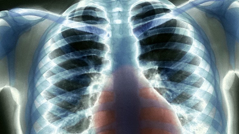
Previously, only X-rays or other such prohibitively expensive methods were capable of tracking medical instruments live during surgical procedures.
Anti-inflammatory drugs ‘reduce cancer deaths by 50%' – study https://t.co/A4n1c0m14Npic.twitter.com/iMmMkR6Cug
— RT (@RT_com) August 30, 2017
The current prototype of the device can track a light source through up to 20cm (7.9 inches) of tissue under normal ambient lighting conditions. "It is now possible to access anatomical regions traditionally out of reach of standard endoscopes" the team wrote in the research paper, published in Biomedical Optics Express on September 1.
"The ability to see a device's location is crucial for many applications in healthcare, as we move forwards with minimally invasive approaches to treating disease," says Kev Dhaliwal, co-author of the research.
“This is an enabling technology that allows us to see through the human body. It has immense potential for diverse applications such as the one described in this work,” Dhaliwal added.
‘Conscious choice’: Swedish hospital refuses to treat failed plastic surgery victimshttps://t.co/sd8m0LqovUpic.twitter.com/9ngPMIXyb9
— RT (@RT_com) August 23, 2017
Currently, the device can detect the position of the fiber optic device inside a human body with a 1cm (0.4 inches) margin of error.
The project is part of the Proteus Interdisciplinary Research Collaboration which specializes in developing technologies for the detection, diagnosis and treatment of lung diseases while preparing for future "robotically augmented surgical procedures."
At present, the camera has been used in surgeries performed on sheep and there is no word yet on when this technology will be tested on human patients.